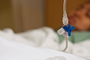
Auscultation an Unsafe Method to Verify Feeding Tube Placement
Call 410-825-5287 for a free medical malpractice consultation

Enteral feeding is a common procedure frequently used to deliver liquid formula and/or medication(s) directly into the stomach of an individual who is unable to consume enough food to meet the needs of their body. Flexible nasogastric tubes, with or without a metal guide, enter through one of the patient's nostrils and must pass the junction with the trachea (airway) before reaching the esophagus (and ultimately the stomach). Since these tubes must move through a treacherous path, serious complications may result if the tube's location is not verified before the initial feeding. Confirmation that any feeding tube has been inserted correctly should be obtained via radiography (x-ray) and one or more recommended supplementary techniques. Unfortunately, auscultation (listening to breathing sounds with a stethoscope) is frequently used in place of acceptable methods, needlessly placing thousands of patients at risk each year.
The Treacherous Path of a Feeding Tube
The human respiratory and digestive systems briefly follow the same path in the section of the throat known as the pharynx. By taking advantage of this shared pathway, feeding tubes can be inserted through a person's nostril and ultimately reach their stomach or small intestine. Unfortunately, feeding tubes are occasionally pushed into the trachea (the windpipe) instead of the esophagus (which leads to the stomach). If the clinician is not aware that the tube is moving "down the wrong pipe", it may be pushed into one of the bronchial tubes and/or lungs where it can cause serious complications including pneumothorax (a collapsed lung). A tube may follow an even more treacherous and lethal path if it is accidentally pushed into a person's cranium and brain. Tubes which were initially inserted correctly can partially slide out of the body between feedings, thereby increasing the risk of choking on liquid formula if the hazard is not noticed by a clinician before the next meal.
Radiology the Most Widely Accepted Method to Verify Correct Tube Placement
The widespread agreement within national guidelines for medical practitioners states that radiography is the most reliable method to ensure correct placement of a feeding tube. Once the tube is believed to have passed the junction of the esophagus and the larynx (the portion of the airway which branches off from the shared pharynx), a chest x-ray should be obtained to ensure it has traversed the correct path. With approval from a radiologist regarding the tube's location, insertion can be completed and a second x-ray obtained to verify that the end has reached the stomach (or in some cases, the small intestine). This entire verification process must be finished before the patient receives his or her first liquid meal and/or medication(s). Radiologists must use extreme vigilance when evaluating chest x-rays of this nature, as tubes which have followed a path into the left lung occasionally appear to have entered the stomach. Patients should also be aware that cumulative exposure to radiation can increase one's chances of cancer and other complications; even so, no other method has proven as reliable for determining the location of a feeding tube.
Techniques Used as Adjuncts to Radiography
When utilized properly, the following procedures/tests may be utilized in conjunction with radiography to reduce the overall number of required x-rays during the initial insertion process. One through three are also considered an acceptable means to verify that the end of a previously inserted feeding tube still remains in the stomach:
- Capnography. Carbon dioxide concentration is an excellent indication of the location of a feeding tube and has been recommended as an alternative to radiography in some studies.
- Measuring the pH of aspirates. When the pH of feeding tube liquid (the aspirate) is between one and five, the clinician can be reasonably certain that the end of the tube is in the stomach.
- Marking the end of the tube. Once a feeding tube has been placed properly it should be marked near the nostril to indicate the total length of material within the body. This mark should be observed before every feeding; if a small portion has slid out, the end has likely risen into the esophagus and the tube should be re-inserted to the initial mark to prevent aspiration of liquid.
- Observing for signs of respiratory distress. A patient who exhibits signs of respiratory distress (coughing or difficulty breathing) during tubal insertion is clearly indicating that the tube has entered his or her airway. Unfortunately, some weak patients or those who are unconscious may not exhibit any such signs; hence, this method can not replace radiography.
Auscultation Widely Used in Place of Acceptable Verification Methods
Despite widespread agreement that auscultation is not a reliable method for determining feeding tube placement, the method remains widely used and in some cases is utilized exclusively. Experts and national guidelines explain that the "whooshing" sound heard through a stethoscope is essentially the same regardless of the location of the end of a feeding tube. Unfortunately, 40 percent of nurses indicated they have not been educated about the unreliability of auscultation, and 88 percent report that they still occasionally use the method in an attempt to check tube location after the initial insertion. Most shocking of all, however, is the fact that auscultation is the only method used to "verify" the correct placement of more than 40 percent of all non-styleted feeding tubes (those which do not contain a metal guide wire). Since as many as 15 percent of all feeding tubes mistakenly pass into the patient's airway during the insertion process, this lack of legitimate location confirmation places thousands of patients at risk of potentially lethal complications each year.
Lack of Education Responsible for This Serious Medical Error
The majority of nurses who acknowledged the use of auscultation to "verify" feeding tube location expressed lack of awareness regarding the national guidelines which explain all acceptable procedures. Clearly, nursing education must improve and hospitals should provide in-service classes to ensure all staff members are kept up to date with important safety guidelines. Patients and their families must also commit to educating themselves and be willing to hold negligent medical care providers responsible for their actions.
Call For a Free Consultation
410-825-5287


Or leave a message...

Office Location:
2300 W. Joppa Rd.
Ste 203
Baltimore, MD 21093
